

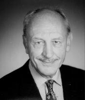
A Symposium in honor of
David R. Davies
Friday, April 25, 1997
National Institutes of Health
Bethesda, MD.
Morning Session (Kiyoshi Mizuchi, chair)
- Brian Matthews University of Oregon - Tolerance and intolerance in protein structure and function
- Alexander Rich Dept of Biology, Massachusetts Institute of Technology - Z DNA and M RNA editing
- Michael Potter Lab of Genetics, National Cancer Institute - Pathogenetic mechanisms in plasmacytoma development
- Gary Felsenfeld Lab of Molecular Biology, NIDDK, NIH - Chromatin structure and gene expression
Afternoon Session (Bill Eaton, chair)
- Edith Miles LBP, NIDDK, NIH - The tryptophan synthase alpha(2)beta(2) complex: architecture, allostery and channeling
- Martin Gellert Lab of Molecular Biology, NIDDK, NIH - New insights into V(D)J recombination
- Robert L. Baldwin Biochemistry Department, Stanford University - Cooperative folding of an apomyoglobin intermediate
- Ira Pastan Lab of Molecular Biology, National Cancer Institute - The design and testing of recombinant immunotoxins in human cancer
- Paul Sigler HHMI/Yale University - Trimeric G proteins: Structure, mechanism and regulation
- Max Perutz MRC, Cambridge, UK - How the structure of proteins was not solved
Symposium Summary
The symposium was opened by Philip Gordon, Director of NIDDK, and the first session was chaired by Kiyoshi Mizuuchi.
Brian Matthews (U. Oregon), a former postdoc of David's, described the work of his laboratory on T-4 lysozyme as a model system for examining protein stability and folding. He likened the structure of T-4 lysozyme to a 'tolerant jigsaw' model. Residues in highly mobile sidechains regions were generally substitutable by other amino acids, while those in the more rigid parts of the molecule were very sensitive to mutation. Insertions into helices caused translocation rather than local bulges. Studies of interior cavities led to recent experiments with rare gases diffused into the protein. Definite difference peaks were observed, with weak peaks indicating either weak binding or fewer electrons. Multiple-methionine substitutions still maintained the three-dimensional structure and activity.
Alex Rich (MIT) , who first invited David to work at the NIH in 1955, spoke about Z-DNA and MRNA editing. Their collaborative work in the early 60s was the first to show regular RNA structures with complementary strands. He discussed Z-forming sequences, which form Z-DNA but are relaxed back to B-form DNA by topoisomerases in a short time. RNA adenosine deaminase, which unwinds double-stranded RNA but not DNA or single-strand RNA, has been extensively studied by his laboratory.
Michael Potter's (NCI) talk followed, entitled 'Pathogenetic mechanisms in plasmacytoma development'. After describing David as the 'Renaissance Man of Structural Biology', he gave a fascinating history of the early discovery and production of monoclonal antibodies. McPc603 was the first myeloma protein with known binding specificity (to phosphocholine) to be structurally characterized. This protein has gained new interest with the recent discovery by Martin Young that the antigen specific for McPC603 is a carbohydrate from Proteus morganii, a bacterium. Recent work by Matthew Scharff and coworkers showed that a single-site mutation in S107, another antiphosphocholine autoantibody, abolishes its phosphocholine specificity but acquires binding to double-stranded DNA.
The morning session concluded with a talk by Gary Felsenfeld, a longtime fellow scientist with David at the Lab of Molecular Biology, NIDDK. His talk, 'Chromatin Structure and Gene Expression' described the simplest chromatin structure as 'beads on a string' separated by linker DNA. The nucleosome beads, 165 bp DNA wrapped around histone octamers, are further compacted into fibers. The transcriptionally active sections are still partially compacted. Promoter and enhancer regions are relaxed and lose the histones, and supercoiling is positive ahead of transcription and negative behind. Recent data shows that the transcribed end reuses the same histone octamer.
After lunch, Bill Eaton (NIDDK) took over as session chair.
The first talk of the afternoon, by Edith Miles (NIDDK), described the structural studies of the tryptophan synthase complex, a collaboration between her lab and David's. Early studies of this complex produced in E.Coli did not crystallize, but (10 years later!) protein from Salmonella produced well-diffracting crystals. The alpha(2)-beta(2) tetramer showed that the active sites in the alpha and beta subunits are 25 A apart, but that a hydrophobic tunnel provides a physical conduit for indole channeling. Recent high resolution studies of mutant enzyme with and without bound ligands to the alpha and beta subunits have provided details about the catalytic and allosteric properties of the enzyme.
Marty Gellert (another founder and current member of LMB, NIDDK) discussed V(D)J recombination. The first step is a site-specific cleavage which generates one hairpinned and one blunt-cut coding end. RAG1 and RAG2 proteins are required, and interestingly, can transfer V(D)J activity to non-lymphoid cells. The cleavage step involves nicking and hairpin formation, for which a divalent ion is required. With Mg++, the reaction stops after the nicking, while with Mn++ it continues to completion.
'Cooperative folding of an apo-myoglobin folding intermediate', was the title of Robert Baldwin's (Stanford) talk. NMR spectra of partially unfolded proteins were shown to have features of both fully folded and fully unfolded states; thus, they were folded in regions. Intriguingly, studies by Schulman and Kim on alpha-lactalbumin have shown exactly the opposite result! Baldwin's group has also looked at mutants involving solvent-exposed residues, and concluded that destabilizing mutations lead to non-cooperative folding. Cooperative folding has been shown to depend on stability and the action of some salts.
Ira Pastan (NCI), another collaborator and tennis partner of David's from NIH, spoke about the design and testing of recombinant immunotoxins in human cancer. The toxin used was Pseudomonas exotoxin A, which kills cells by interfering with the ribosomal machinery. The second and third domains of this toxin were attached via a lysine to the B3 antibody, whose antigen is a carbohydrate is highly expressed in many cancers. This immunotoxin was put into clinical trials with significant success, but it was discovered to have a long half-life and to cause damage to epithelial cells. A refined version had the variable domains of the antibody as a single chain, and had a shorter half-life but was susceptible to aggregation. A disulfide-stabilized Fv made by mutating two residues to Cys was much more stable and is currently in clinical trials.
Paul Sigler (Yale) described his learning experience as a postdoc in David's lab, and went on to discuss 'Trimeric G proteins: Structure, mechanism and regulation'. He described G-protein mediated transmembrane signalling, focusing on transducin and its signalling action in the retina. The alpha subunit of this 3-domain protein has been solved in complex with a GTP analog, GDP and GDP+AlF(4). The beta and gamma subunits were solved independently, as well as in complex with GDP and a GTP analog. This family of structures showed that the GTP-GDP change was accompanied by three loop movements, and identified a possible membrane-binding surface.
Returning to the origins of protein crystallography, Max Perutz closed the symposium with 'How the structure of proteins was not solved'. He described the intellectual excitement at Cambridge starting in the 30s, with photographs of John Bernal, Dorothy Crowfoot (later Hodgkin) and W.L. Bragg in those days. During the early days of his work on hemoglobin, solving the structure of a protein was considered a hopeless task by many crystallographers. He traced the breakthroughs in the diffraction analysis, mostly done by hand and eye in those days before computers were commonly available. Once it was determined that a heavy atom crystallized along with the protein could be detected in the diffraction pattern, the structure was deemed solved. (Michael Rossman followed this talk with an anecdote: at the time he was considering working with Perutz, another crystallographer told him 'You know, what Max does is not really science'!)
The symposium was followed by a banquet at the NIH Cloisters, enlivened by reminescences from Brian Matthews, Paul Sigler, Alex Wlodawer, Rick Bott, Sally Davies, Gary Felsenfeld, and Marty Gellert.
-- Susan Chacko